Overview
A scan is not a single event. It starts with a clinician's question and ends, if all goes well, with an answer that changes the patient's care. In between, it passes through many people and several computer systems. This lesson follows Mr. Diaz's CT pulmonary angiogram (CTPA) from start to finish. Module 3 then shows where AI tools plug into this same path. About 15-20 minutes. New terms are in the Module 2 glossary (opens in a new tab).
By the end of this lesson you can:
- Describe the nine steps of a scan's journey, from order to follow-up.
- Name the main computer systems (EHR, RIS, PACS) and what each one does.
- Explain why the reason for the exam on the order matters so much.
- Recognize the steps where errors and delays most often happen.
- State the two rules that close the loop: critical results need a direct call, and follow-up recommendations need an owner.
Radiology informatics 101: how radiology data travels
Radiology informatics is the study and running of the computer systems that move radiology information: orders, images, reports and results. You will hear these names on your first day in any hospital. Each is a separate piece of software, often from a different company, and they pass messages to each other.
These systems talk using shared standards. DICOM carries images (more in Lesson 2.3). HL7 (Health Level Seven) carries text messages such as orders and reports. FHIR (Fast Healthcare Interoperability Resources, said "fire") is a newer, web-style version of HL7 that many apps, and many AI tools, now use to read from the EHR.
Put together, one scan's data travels in a loop. The order starts in the EHR and goes to the RIS as an HL7 message. The RIS books the exam and sends the patient's details to the scanner's own worklist, so the images are labeled with the right name and exam. The scanner sends the images to PACS as DICOM files, and the exam appears on the radiologist's worklist. The radiologist reads it in PACS, dictates the report in the reporting system, and the signed report travels back through the RIS to the EHR, where the ordering clinician and, often, the patient read it. A break at any link (a wrong name on the order, images that never reach PACS, a report that never reaches the chart) is a break in patient care.
Follow Mr. Diaz's scan
Step through the journey. The map shows which system is involved at each step.
Why the order matters
The reason for exam is the radiologist's only window into the patient. They usually have not met the patient and see the order long before the full chart. A good reason for exam changes how the scan is done (the protocol) and what the radiologist looks for.
| Weak reason for exam | Better reason for exam |
|---|---|
| "Chest pain" | "Sudden right pleuritic chest pain and shortness of breath after a long flight; D-dimer elevated. Assess for PE." |
| "Rule out pathology" | "Right lower abdominal pain for 1 day, fever, white count 15. Appendicitis?" |
| "Headache" | "Sudden worst headache of life 2 hours ago, on blood thinners. Bleed?" |
The ACR Appropriateness Criteria are guidelines from the American College of Radiology that rate which test is best for a given clinical problem. They are what the decision support in step 1 is usually built on.
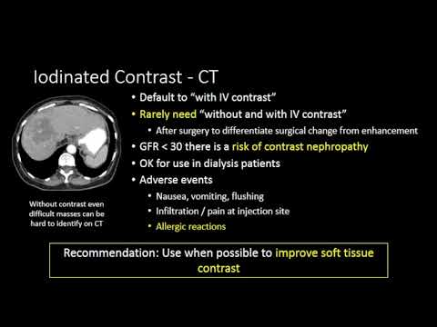

Put it in order
Drag the steps into the order they happen. Then check. (With a keyboard: focus a step and use the up and down arrows.)
- The radiologist reads the images and compares with priors
- The exam is protocolled and scheduled in the RIS
- The radiologist phones the team about the critical result, then signs the report
- The images are sent to PACS and matched to the order
- The clinician enters an order with a reason for exam
- A follow-up CT is booked for the lung nodule
- The exam appears on the worklist, marked STAT
- The technologist checks identity and runs the scan
- The radiologist dictates the report
Where things go wrong
Most imaging errors are not a radiologist missing something on the picture. Many happen before the scan or after the report. Knowing the steps helps you see where.
Each story is a real type of error. Decide at which step it happened.
Two rules that close the loop
Two of the stories above failed after the radiologist had done the read correctly. Both failures have a simple rule against them.
A finding that needs action now, such as a PE, a collapsed lung or a bleed in the brain, is never left to sit in the chart. The radiologist phones the treating clinician, the clinician repeats it back ("read-back"), and the call is written into the report: who, when and what was said. If the ordering doctor cannot be reached, the radiologist keeps going up the chain until someone who can act has been told.
A recommendation in a report does nothing on its own. A named person has to tell the patient, order the next scan or hand the task over clearly, and put it in the discharge summary. By default that is the ordering physician's team. Tracking programs are a safety net, not a replacement for an owner.
Three habits help most: write a real reason for exam, read the whole report (not only the first line), and when a report recommends follow-up, make sure someone owns it before the patient leaves. Make sure the radiology department can reach you, or whoever covers for you, when a critical result comes in.
Where AI fits (a preview)
Every step on the map is a place a computer could help, and AI tools already exist for most of them: suggesting a protocol, cleaning up noisy images, moving suspected critical findings to the top of the worklist, flagging findings during the read, drafting parts of the report, and tracking follow-up recommendations. Each one changes the journey in a different way, and each can fail in a different way. Lesson 3.1 maps these tools onto the journey you just learned.
Knowledge check
Answer each question to continue. Any answer is fine; read the explanation.
Which system stores the images and shows them on the radiologist's workstation?
PACS (picture archiving and communication system) stores and displays images. The EHR is the patient's chart; the RIS schedules and tracks radiology exams.
The radiologist finds Mr. Diaz's PE at 2 a.m. What should happen besides signing the report?
A PE is a critical result. It needs direct, documented communication so treatment starts right away.
Which step of the journey is most often where an incidental finding gets lost?
Recommendations for later imaging depend on someone reading, owning and scheduling them. Handoffs between teams make this the weakest step.
Key takeaways
- A scan's journey has nine steps: order, protocol and schedule, scan, store and send, worklist, read, report, communicate, follow-up.
- The EHR holds the chart, the RIS schedules and tracks, PACS stores and shows images. They talk through DICOM, HL7 and FHIR.
- The reason for exam shapes both how the scan is done and what the radiologist looks for.
- Critical results need a direct call. Follow-up recommendations need an owner.
- Many errors happen before the scan or after the report, not only during the read.
Go further (optional)
- ACR Appropriateness Criteria (look up "suspected pulmonary embolism" and see how the tests are rated)
- How to Order Diagnostic Imaging (Joseph W. Owen, MD, YouTube)
Next lesson: the report is signed. What does it say, and how do you read it?